Sarcoma PanelSarcomas are representing a heterogeneous group of tumors originating from mesenchymal tissue. Around 80% develop from soft tissue, the remaining 20% from bone. Sarcomas are frequently observed in children and young adults, accounting for more than 20% of all pediatric neoplasms. Due to the diversity, more than 100 subtypes are known to date, the diagnosis of sarcomas is challenging and fluorescence in situ hybridization has become a valuable tool to analyze the molecular signature of sarcoma subtypes today. We are very proud to introduce a probe panel covering a subset of the most relevant chromosomal loci associated with the development of sarcomas. All probes are intended for hybridization on methanol/acetic-acid fixed cells and formalin-fixed, paraffin-embedded (FFPE) tissue sections. In combination with our optimized TissueFISH Pretreatment Kit, this panel will make your workflow more efficient.
|
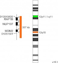
XL MDM2D-5047-100-OG Clinical Applications: Solid tumorsThe XL MDM2 hybridizes with an orange labeled probe to MDM2 and its flanking regions. A green labeled probe hybridizes to the centromere of chromosome 12. MDM2 is an ubiquitin ligase negatively regulating p53. MDM2 is amplified in about 7% of all human cancers with the highest frequency of about 20% in soft tissue tumors. The analysis of the MDM2 amplification status by FISH is considered as a useful technique for the differential diagnosis of well-differentiated liposarcomas/atypical lipomatous tumors and benign lipomatous tumors since benign lesions do not harbor MDM2 amplifications. Download Fact Sheet (PDF)
|
 |
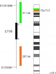
XL ETV6D-5073-100-OG Clinical Applications: Solid tumorsThe XL ETV6 probe is designed as a break apart probe. Its orange labeled part hybridizes proximal to the ETV6 (TEL) gene at 12p13, the green labeled probe hybridizes to the distal region of ETV6. The ETV6 gene, located on 12p13.2, is involved in numerous translocations found in myeloid and lymphoid malignancies. Besides hematological malignancies, ETV6 is also involved in the development of the pediatric congenital mesoblastic nephroma and fibrosarcoma. t(12;15)(p13;q25) fuses ETV6 to the tyrosin receptor kinase NTRK3 resulting in the ETV6-NTRK3 fusion gene, a constitutively active tyrosine kinase. Download Fact Sheet (PDF)
|
|
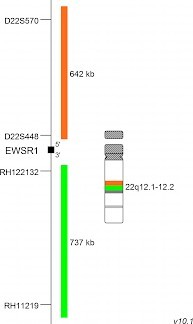
XL EWSR1 BAD-6011-100-OG Clinical Applications: Solid tumorsXL EWSR1 BA is designed as a break apart probe. The orange labeled probe hybridizes proximal to the breakpoint in the EWSR1 gene region at 22q12, the green labeled probe hybridizes distal to the breakpoint. The Ewing sarcoma (EWS) is a rare and highly aggressive cancer with an incidence of about three in one millions Caucasians. EWS is typified by chromosomal translocations resulting in fusion genes between the EWS RNA Binding Protein 1 (EWSR1) and a member of the group of ETS transcription factors. t(11;22)(q24;q12) is the most common of these translocations represented by the EWSR1-FLI1 fusion gene with a frequency of about 85%. Download Fact Sheet (PDF)
|
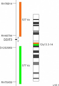
XL DDIT3 BAD-6032-100-OG Clinical Applications: Solid tumorsXL DDIT3 BA is designed as a break apart probe. The orange labeled probe hybridizes proximal to the breakpoint in the DDIT3 gene region at 12q13, the green labeled probe hybridizes distal to the breakpoint. The myxoid/round cell liposarcoma (MRCLS) is the most common liposarcoma with an increased incidence in the extremities. It is characterized by the reciprocal translocation t(12;16)(q13;p11) which is present in up to 95% of cases. The resulting FUS-DDIT3 fusion protein has oncogenic potential and interferes with adipogenic differentiation. EWSR1 is an alternative translocation partner of DDIT3 and the resulting fusion protein EWSR1-DDIT3, originating from t(12;22)(q13;q12), accounts for <5% of MRCLS cases. Download Fact Sheet (PDF)
|
|
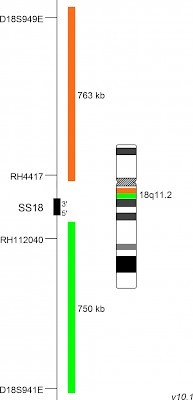
XL SS18 BAD-6033-100-OG Clinical Applications: Solid tumorsXL SS18 BA is designed as a break apart probe. The orange labeled probe hybridizes proximal to the breakpoint in the SS18 gene region at 18q11, the green labeled probe hybridizes distal to the breakpoint. The synovial sarcoma is a highly aggressive and relatively rare soft tissue sarcoma. It often develops in the limbs of adolescents and young adults and comprises about 10-20% of soft tissue sarcomas in this population. The disease is characterized by the balanced translocation t(X;18) resulting in an in-frame fusion of ´synovial sarcoma translocation, chromosome 18´ (SS18) with members of the ´synovial sarcoma, X breakpoint´ family (SSX), located on the X chromosome. Download Fact Sheet (PDF)
|
|
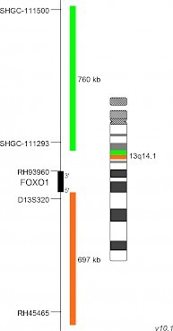
XL FOXO1 BAD-6034-100-OG Clinical Applications: Solid tumorsXL FOXO1 BA is designed as a break apart probe. The green labeled probe hybridizes proximal to the breakpoint in the FOXO1 gene region at 13q14, the orange labeled probe hybridizes distal to the breakpoint. Rhabdomyosarcoma is a relatively rare cancer type but it is the most common soft tissue sarcoma in children and adolescents. The histopathological classification includes several subtypes. The alveolar rhabdomyosarcoma is associated with a worse outcome and is characterized by the two reciprocal translocations t(2;13)(q35;q14) and t(1;13) (p36;q14), affecting the FOXO1 gene region and PAX3 or PAX7 respectively, in about 80% of cases. Download Fact Sheet (PDF)
|
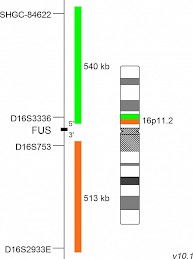
XL FUS BAD-6035-100-OG Clinical Applications: Solid tumorsXL FUS BA is designed as a break apart probe. The orange labeled probe hybridizes proximal to the breakpoint in the FUS gene region at 16p11, the green labeled probe hybridizes distal to the breakpoint. Myxoid liposarcomas (MLS) are accounting for about 30% of liposarcomas and represent approximately 10% of adult soft tissue sarcomas. The most common aberration in MLS is the translocation t(12;16)(q13;p11) with a frequency of about 95% and to a much lesser extend t(12;22)(q13;q12), in which FUS is not involved. These reciprocal translocations are resulting in the generation of FUS-DDIT3 and EWSR1-DDIT3 fusion genes, respectively. Download Fact Sheet (PDF)
|
|
TissueFISH Pretreatment KitD-0905-025-TF Clinical Applications: Solid tumorsThe MetaSystems' TissueFISH Pre-treatment Kit contains reagents and solutions to process formalin-fixed, paraffin-embedded tissue sections for FISH assays. Reagents provided are: 5x 5 ml 10x Pretreatment Buffer, 200 ml Protease Buffer, 500 µl Protease Stock Solution.
|
|
DAPI/antifade, 500µlD-0902-500-DA Chromosome counterstain in a buffer containing anti-photobleaching (antifade) reagent. 250ng/ml, 500µl
|