XL MYCN amp
Amplification Probe
- Order Number
- D-6031-100-OG
- Package Size
- 100 µl (10 Tests)
- Chromosome
- 022
- Regulatory Status
- IVDD
IVDR Certification
MetaSystems Probes has already certified a wide range of FISH probes, according to IVDR.
This product remains IVDD-certified until further notice.
Discover all IVDR-certified products
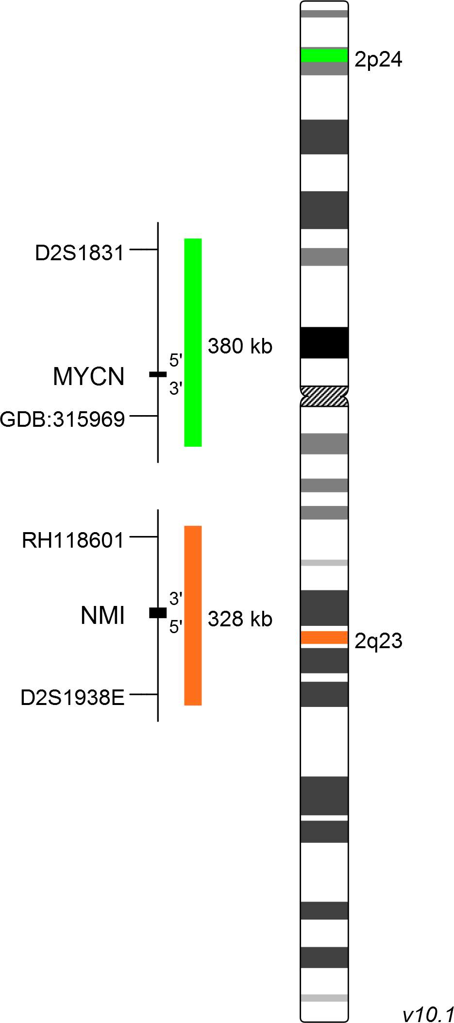
XL MYCN amp consists of a green-labeled probe hybridizing to the MYCN gene region at 2p24 and an orange-labeled probe hybridizing to the NMI gene region at 2q23.
Probe maps for selected products have been updated. These updates ensure a consistent presentation of all gaps larger than 10 kb including adjustments to markers, genes, and related elements. This update does not affect the device characteristics or product composition. Please refer to the list to find out which products now include updated probe maps.
Probe map details are based on UCSC Genome Browser GRCh37/hg19, with map components not to scale.
The MYCN gene is a member of the MYC transcription factor family consisting of c-MYC, MYCN and MYCL. It is crucial for embryonic development, especially in the nervous system. The expression is tightly regulated in a spatial and timely manner and high levels are found in the developing brain, retina, neuroepithelial cells, lung and kidney. MYCN expression is associated with the maintenance of self-renewal capacity and the pluripotent status of stem cells. Dysregulation of MYCN contributes to the development of different kinds of childhood tumors including neuroblastoma, medulloblastoma, rhabdomyosarcoma and Wilms tumor. MYCN is also involved in the development of some adulthood neoplasms such as prostate and lung cancer.
Neuroblastoma is the most common extracranial solid tumor in infants. About 6% of all cancers in children are caused by neuroblastomas and 20-25% of neuroblastoma patients are showing an amplification of the MYCN gene. MYCN amplification is an important prognostic marker for risk stratification in neuroblastoma. Generally, patients with MYCN amplification have a poor prognosis. FISH is a valuable tool for the analysis of the MYCN amplification status. It detects MYCN amplification on single-cell level and allows the correlation with morphological cell features.
Clinical Applications
- Solid Tumors (Solid Tumors)

Normal Cell:
Two green (2G) and two orange (2O) signals.

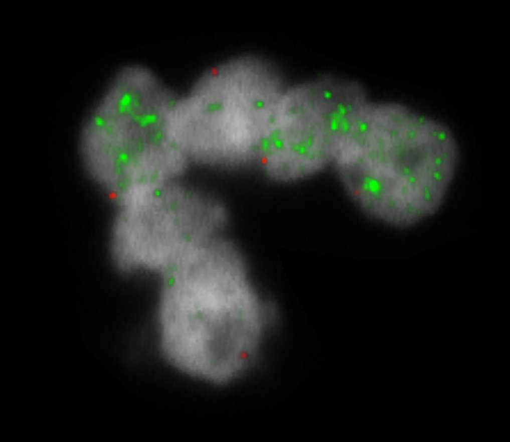
Aberrant Cell (typical results):
Two orange (2O), one green (1G) and green signal clusters resulting from an amplification of the green signal (homogenously staining region).

Aberrant Cell (typical results):
Two orange (2O) and multiple green (mG) signals resulting from amplification of the green signal (double minutes).
- Theissen et al (2009) Clin Cancer Res 15:2085-2090
- Huang and Weiss (2013) Cold Spring Harb Perspect Med 2013;3(10):a014415
- Ruiz-Pérez et al (2017) Genes 8(4):113
Certificate of Analysis (CoA)
or go to CoA Database


