XL EGFR amp
Amplification Probe
- Order Number
- D-6005-100-OG
- Package Size
- 100 µl (10 Tests)
- Chromosome
- 077
- Regulatory Status
- IVDD
IVDR Certification
MetaSystems Probes has already certified a wide range of FISH probes, according to IVDR.
This product remains IVDD-certified until further notice.
Discover all IVDR-certified products
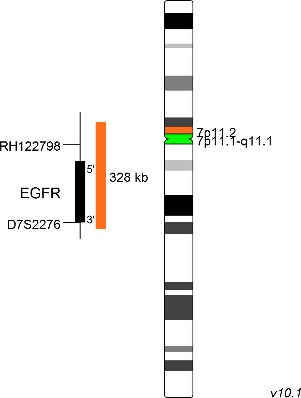
XL EGFR amp consists of an orange-labeled probe hybridizing to the EGFR gene region at 7p11.2 and a green-labeled probe hybridizing to the centromere of chromosome 7.
Probe maps for selected products have been updated. These updates ensure a consistent presentation of all gaps larger than 10 kb including adjustments to markers, genes, and related elements. This update does not affect the device characteristics or product composition. Please refer to the list to find out which products now include updated probe maps.
Probe map details are based on UCSC Genome Browser GRCh37/hg19, with map components not to scale.
EGFR (epidermal growth factor receptor) gene amplification generally results in increased protein expression in breast carcinomas. About 6% of breast carcinomas show moderate to low-level EGFR amplification associated with genuine EGFR protein overexpression. Studies in non-small cell lung cancer (NSCLC) have shown that EGFR expression is associated with reduced survival, frequent lymph node metastasis, and poor chemosensitivity.
EGFR is a member of the ErbB family of receptors, a subfamily of four closely related receptor tyrosine kinases which all play an important role in controlling normal cell growth, apoptosis, and other cellular functions. Mutations of EGFRs can lead to NSCLC, pancreatic cancer, breast cancer, colon cancer, and some other cancers.
New drugs such as gefitinib and erlotinib directly target the EGFR. EGFR-positive patients have shown a 60% response rate, which exceeds the response rate for conventional chemotherapy.
Clinical Applications
- Solid Tumors (Solid Tumors)

Normal Cell:
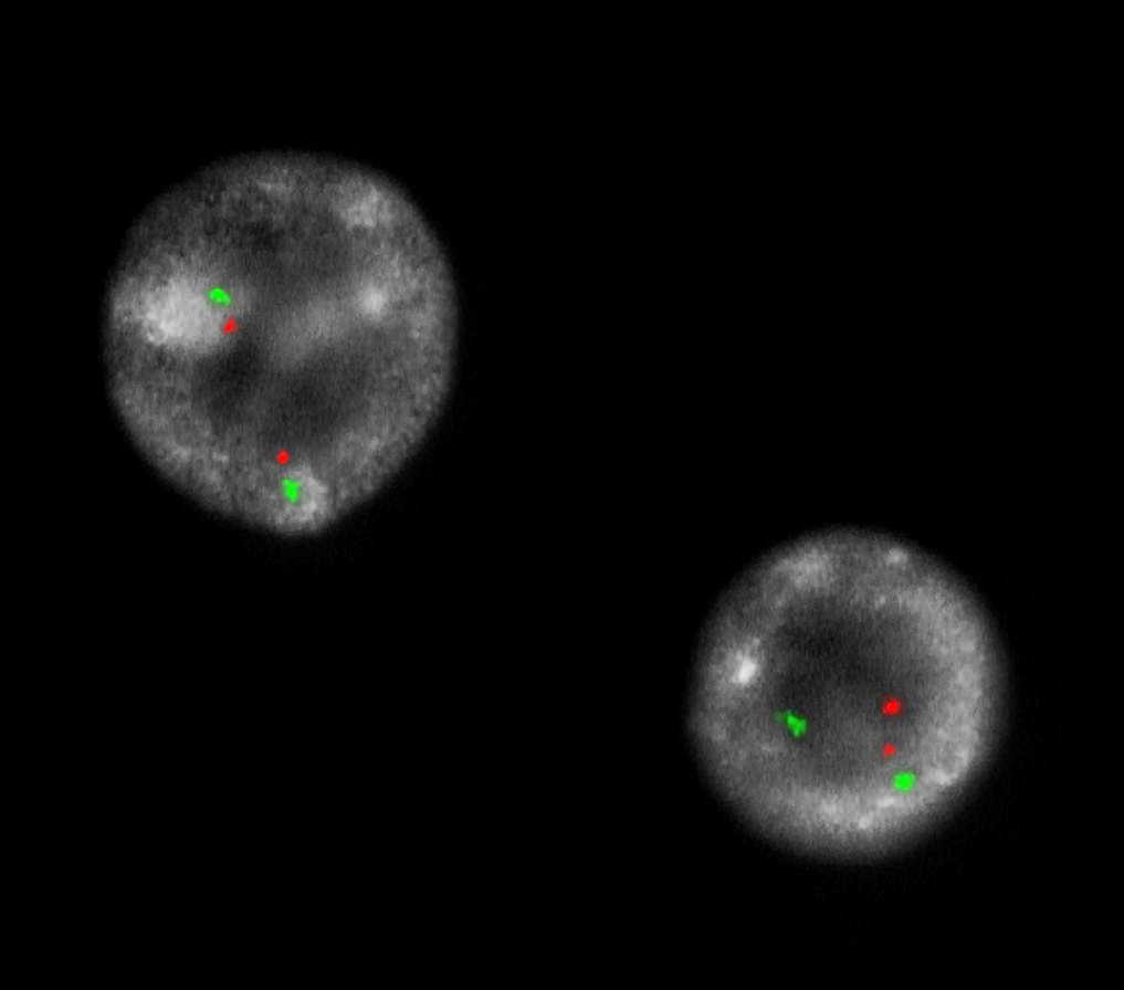
Two green (2G) and two orange (2O) signals.

Aberrant Cell (typical results):
Two green (2G) and one separate orange (1O) signal, and orange signal clusters indicating amplification of EGFR (homogeneously staining region =HSR).

Aberrant Cell (typical results):
Two green (2G) and multiple copies of orange signals indicating amplification of EGFR (double minute=dm).
- Okada et al (2003) Cancer Res 63:413-416
- Bhargava et al (2006) Mod Patho 18:1027-1033
- Sholl et al (2009) Cancer Res 69:8341-8348
Certificate of Analysis (CoA)
or go to CoA Database


