XL MYC amp
Amplification Probe
- Order Number
- D-6008-100-OG
- Package Size
- 100 µl (10 Tests)
- Chromosome
- 088
- Regulatory Status
- IVDD
IVDR Certification
MetaSystems Probes has already certified a wide range of FISH probes, according to IVDR.
This product remains IVDD-certified until further notice.
Discover all IVDR-certified products
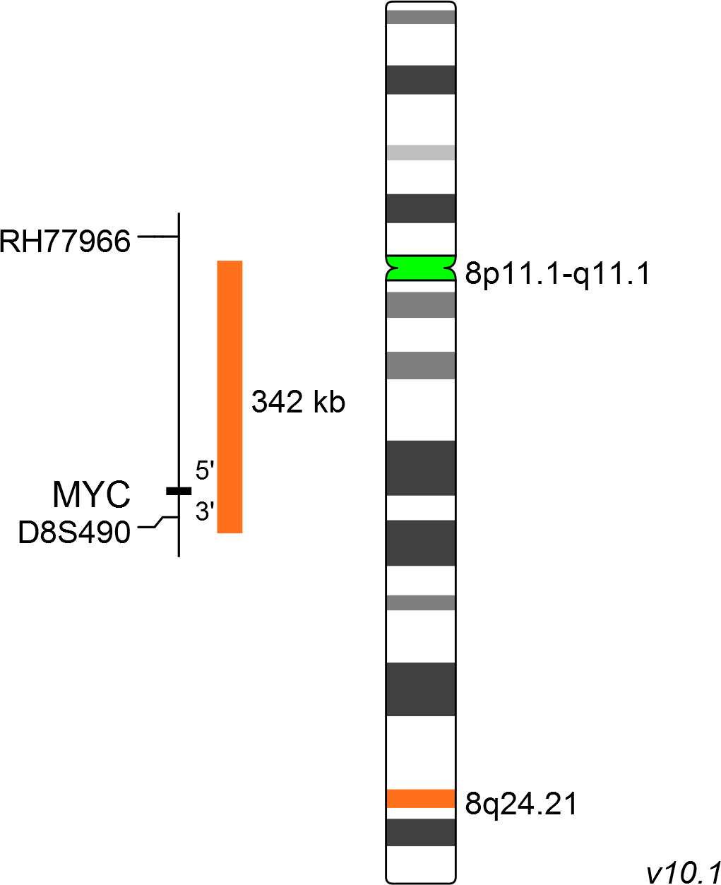
XL MYC amp consists of an orange-labeled probe hybridizing to the MYC gene region at 8q24.21 and a green-labeled probe hybridizing to the centromere of chromosome 8.
Probe maps for selected products have been updated. These updates ensure a consistent presentation of all gaps larger than 10 kb including adjustments to markers, genes, and related elements. This update does not affect the device characteristics or product composition. Please refer to the list to find out which products now include updated probe maps.
Probe map details are based on UCSC Genome Browser GRCh37/hg19, with map components not to scale.
Amplification of MYC has been described in many types of tumor, including breast, cervical and colon cancers, as well as in squamous cell carcinomas of the head and neck, myeloma, non-Hodgkin lymphoma, gastric adenocarcinomas and ovarian cancer. MYC is the most frequently amplified oncogene and the elevated expression of its gene product correlates with tumor aggression and poor clinical outcome.
The proto-oncogene MYC, located at 8q24.1, encodes a nuclear phosphoprotein transcription factor which has an integral role in a variety of cellular processes, such as cell cycle progression, proliferation, metabolism, adhesion, differentiation, and apoptosis.
Clinical Applications
- Solid Tumors (Solid Tumors)

Normal Cell:
Two green (2G) and two orange (2O) signals.

Aberrant Cell (typical results):
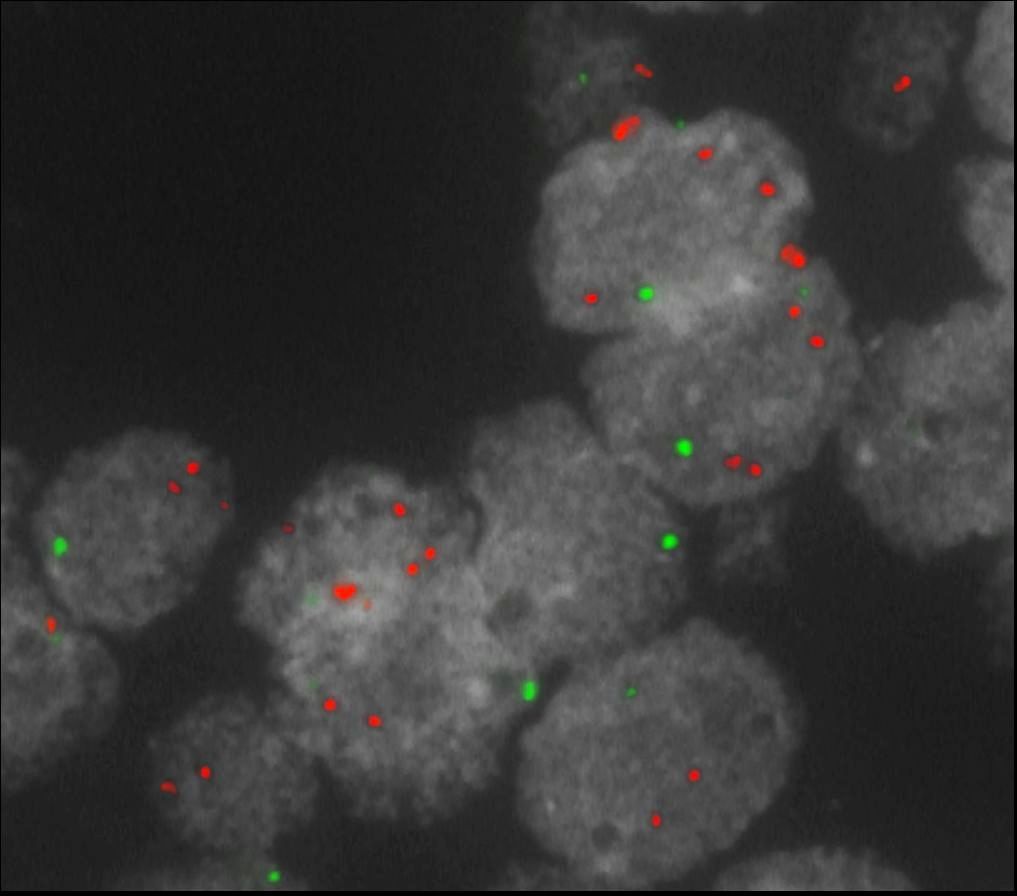
Two green (2G) and one separate orange (1O) signal, and orange signal clusters indicating amplification of MYC (homogeneously staining region = HSR).

Aberrant Cell (typical results):
Two green (2G) and multiple copies of orange signals indicating amplification of MYC (double minute=dm).
- Rummukainen et al (2001) Mod Pathol 14:1030-1035
- Blancato et al (2004) Br J Cancer 90:1612-1619
- Singhi et al (2012) Mod Pathol 25:378-387
Certificate of Analysis (CoA)
or go to CoA Database


