XL DDIT3 BA
Break Apart Probe
- Order Number
- D-6032-100-OG
- Package Size
- 100 µl (10 Tests)
- Chromosome
- 1212
- Regulatory Status
- IVDD
IVDR Certification
MetaSystems Probes has already certified a wide range of FISH probes, according to IVDR.
This product remains IVDD-certified until further notice.
Discover all IVDR-certified products
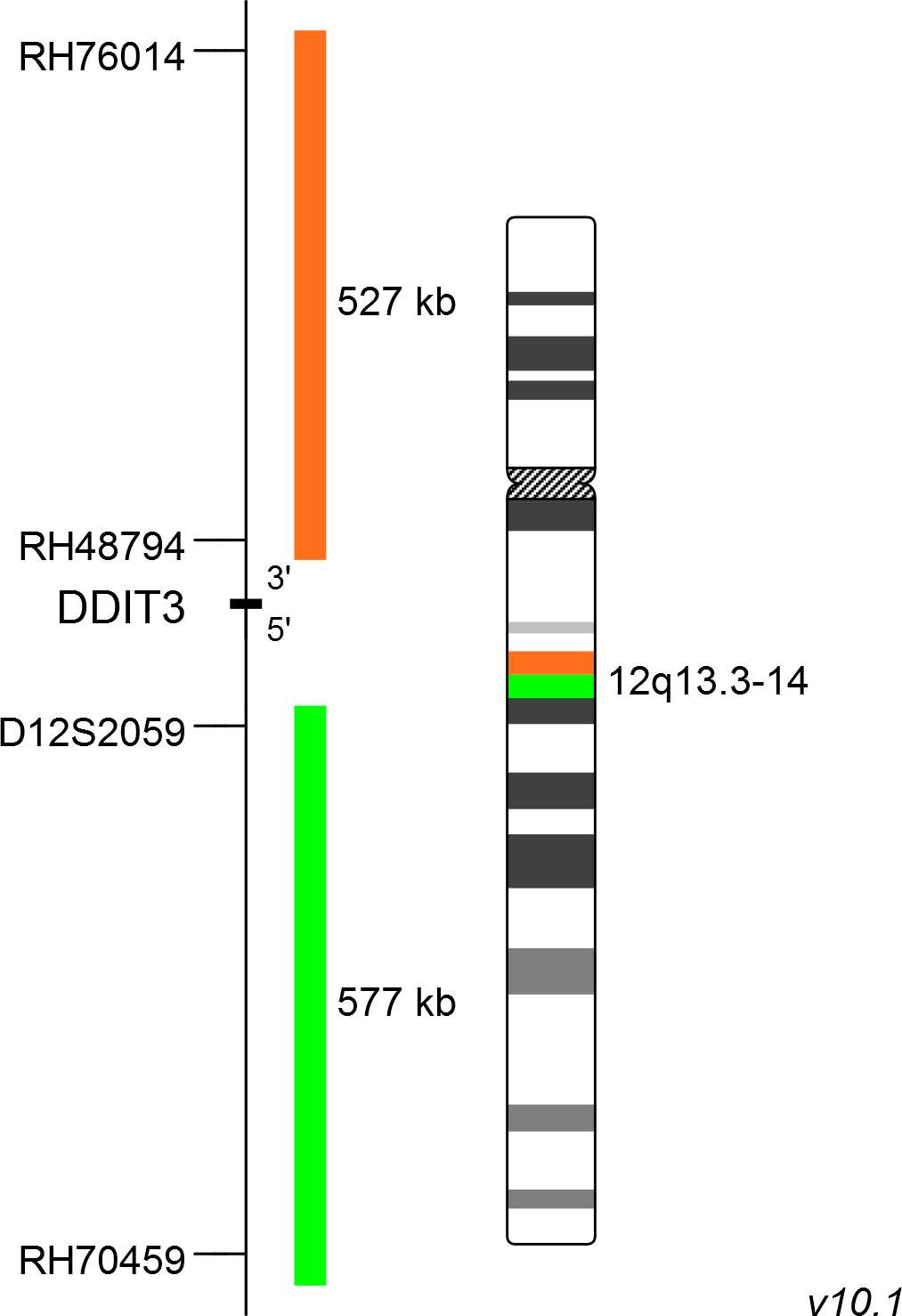
XL DDIT3 BA consists of an orange-labeled probe hybridizing proximal to the DDIT3 gene region at 12q13.3 and a green-labeled probe hybridizing distal to the DDIT3 gene region at 12q13.3-14.
Probe maps for selected products have been updated. These updates ensure a consistent presentation of all gaps larger than 10 kb including adjustments to markers, genes, and related elements. This update does not affect the device characteristics or product composition. Please refer to the list to find out which products now include updated probe maps.
Probe map details are based on UCSC Genome Browser GRCh37/hg19, with map components not to scale.
Histological similarities and a subset of common immunohistochemical features make the diagnosis of soft tissue neoplasms challenging. Several entities characterizing reciprocal translocations have been identified and FISH based assays specific for the detection of these aberrations have become a valuable tool in diagnostics today. Myxoid/round cell liposarcoma (MRCLS) is the most common liposarcoma with an increased incidence in the extremities. Compared to other liposarcomas, MRCLS has a greater tendency for metastasis. It is characterized by the reciprocal translocation t(12;16)(q13;p11) which is present in up to 95% of cases. The resulting FUS-DDIT3 fusion protein has oncogenic potential and interferes with adipogenic differentiation. A paralog of FUS with high sequence homology is the EWS RNA Binding Protein 1 (EWSR1). EWSR1 is an alternative translocation partner of DDIT3 and the resulting fusion protein EWSR1-DDIT3, originating from t(12;22)(q13;q12), accounts for <5% of MRCLS cases. DDIT3 rearrangements are highly specific for MRCLS due to the presence of FUS-DDIT3 or EWSR1-DDIT3 fusion genes.
Clinical Applications
- Solid Tumors (Solid Tumors)

Normal Cell:
Two green-orange colocalization/fusion signals (2GO).

Aberrant Cell (typical results):
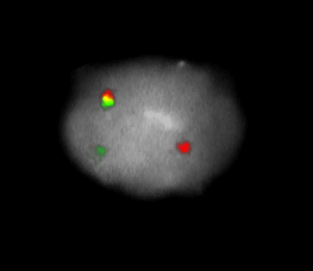
One green-orange colocalization/fusion signal (1GO), one separate green (1G) and orange (1O) signal each resulting from a chromosome break in the relevant locus.
- Cristina et al (2000) JMD 2:132-138
- Downs-Kelly et al (2008) Am J Surg Pathol 32:8-13
- Tanas et al (2009) Adv Anat Pathol 16:383-391
Certificate of Analysis (CoA)
or go to CoA Database


