XA TriScore (X/Y/21)
Aneusomy Probe
- Order Number
- D-5603-100-TC
- Package Size
- 100 µl (10 Tests)
- Regulatory Status
- IVDR
IVDR Certification

This probe is IVDR-certified in compliance with the Regulation (EU) 2017/746 for in vitro diagnostic medical devices (IVDR).
MetaSystems Probes has already certified a wide range of FISH probes, according to IVDR.
This product may not be available in all countries outside the European Union.

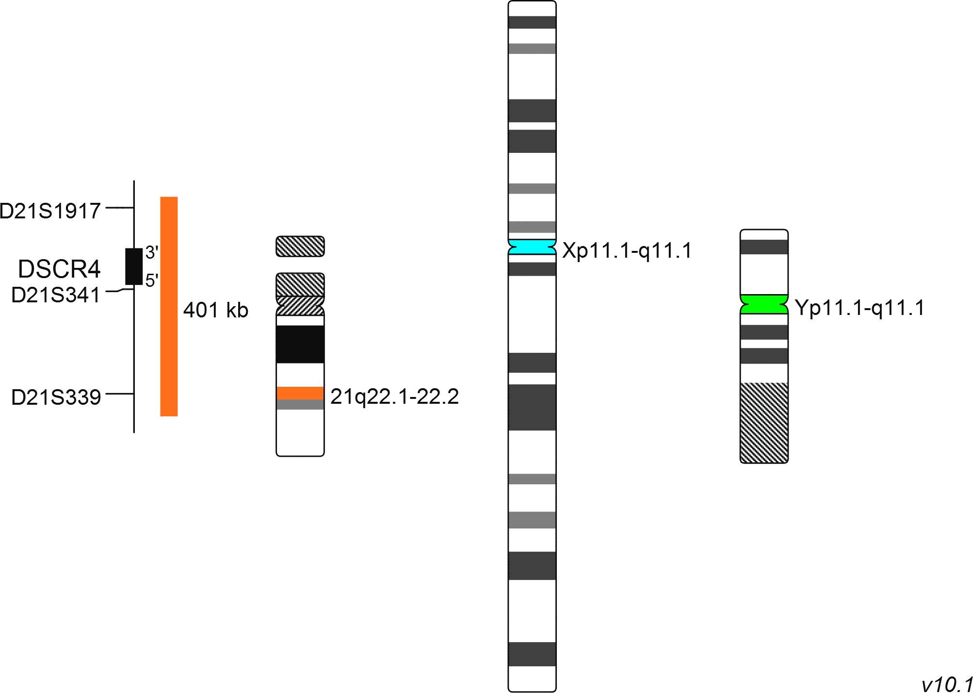
The XA TriScore (X/Y/21) enumeration probe consists of an aqua-labeled probe hybridizing to the centromere of the X-chromosome, a green-labeled probe hybridizing to the centromere of the Y-chromosome and an orange-labeled probe hybridizing to the DSCR4 gene region at 21q22.1-22.2.
Probe maps for selected products have been updated. These updates ensure a consistent presentation of all gaps larger than 10 kb including adjustments to markers, genes, and related elements. This update does not affect the device characteristics or product composition. Please refer to the list to find out which products now include updated probe maps.
Probe map details are based on UCSC Genome Browser GRCh37/hg19, with map components not to scale.

Normal Cell (male):
One green (1G), two orange (2O), and one blue (1B) signal.

Normal Cell (female):
No green (0G), two orange (2O), and two blue (2B) signals.

Aberrant Cell (male):
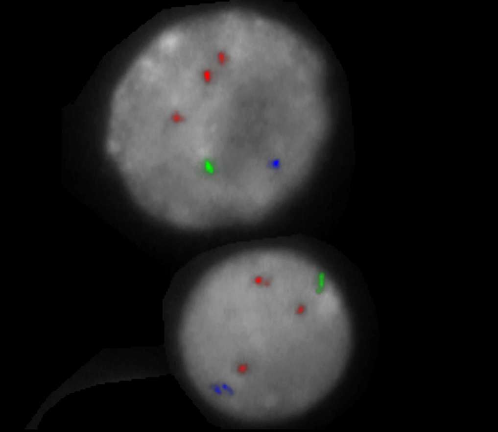
One green (1G), three orange (3O), and one blue (1B) signal.

Aberrant Cell (female):
No green (0G), three orange (3O), and two blue (2B) signals.

Aberrant Cell (male):
One green (1G), two orange (2O), and two blue (2B) signals.

Aberrant Cell (male):
Two green (2G), two orange (2O), and two blue (2B) signals.

Aberrant Cell (female):
No green (0G), two orange (2O), and one blue (1B) signals.

Aberrant Cell (female):
No green (0G), two orange (2O), and three blue (3B) signals.


